Human Mitochondrial Antiviral Signaling Protein (MAVS) Protein
208€ (10 µg)
Por favor contáctenos para obtener información detallada sobre el precio y disponibilidad.
Name
Human Mitochondrial Antiviral Signaling Protein (MAVS) Protein
Category
Proteins and Peptides
Provider
Abbexa
Reference
abx655487
Tested Applications
WB, SDS-PAGE
Description
Human Mitochondrial Antiviral Signaling Protein (MAVS) Protein is a recombinant Human protein expressed in E. coli.
Documentos del producto
Instrucciones
Data sheet
Especificaciones del producto
| Category | Proteins and Peptides |
| Immunogen Target | Mitochondrial Antiviral Signaling Protein (MAVS) |
| Host | E. coli |
| Assay Type | Activity: Not tested Sequence Fragment: Met1-Gly400 Tag: His and TRxA Tag |
| Origin | Human |
| Conjugation | Unconjugated |
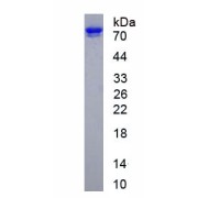
| Observed MW | Calculated MW: 62.0 kDa Observed MW: 75 kDa |
| Expression | Recombinant |
| Purity | > 90% |
| Size 1 | 10 µg |
| Size 2 | 50 µg |
| Size 3 | 100 µg |
| Size 4 | 200 µg |
| Size 5 | 500 µg |
| Form | Lyophilized |
| Tested Applications | WB, SDS-PAGE |
| Buffer | Prior to lyophilization: PBS, pH 7.4, containing 0.01% Sarcosyl, 1 mM DTT, 5% Trehalose and Proclin-300. |
| Availability | Shipped within 5-7 working days. |
| Storage | Store lyophilized form at 2-8°C for up to 1 month. For longer periods, store lyophilized or liquid at -80°C. Avoid repeated freeze–thaw cycles. |
| Dry Ice | No |
| Background | Protein MAVS |
| Status | RUO |
| Note | THIS PRODUCT IS FOR RESEARCH USE ONLY. NOT FOR USE IN DIAGNOSTIC, THERAPEUTIC OR COSMETIC PROCEDURES. NOT FOR HUMAN OR ANIMAL CONSUMPTION. To keep the original salt concentration, we recommend reconstituting to the original concentration prior to lyophilization (see Concentration) in ddH2O. If a lower concentration is required, dilute in PBS, pH 7.4. If a higher concentration is required, the product can be reconstituted directly in PBS, pH 7.4, though please note that this will change the overall salt concentration. The stock concentration should be between 0.1-1.0 mg/ml. Do not vortex. Concentration: Prior to lyophilization: 200 µg/ml |
Productos relacionados

Mitochondrial Antiviral Signaling Protein (MAVS) Antibody
MAVS Antibody is a Rabbit Polyclonal antibody against MAVS. This gene encodes an intermediary protein necessary in the v…
Ver producto
Mitochondrial Antiviral Signaling Protein (MAVS) Antibody
Double-stranded RNA viruses are recognized in a cell type-dependent manner by the transmembrane receptor TLR3 (MIM 60302…
Ver producto
Mitochondrial Antiviral Signaling Protein (MAVS) Antibody
Mitochondrial Antiviral Signaling Protein Antibody is a Rabbit Polyclonal antibody against Mitochondrial Antiviral Signa…
Ver producto